UOC Radiologia ASST Bergamo Est Direttore Dr Gianluigi Patelli
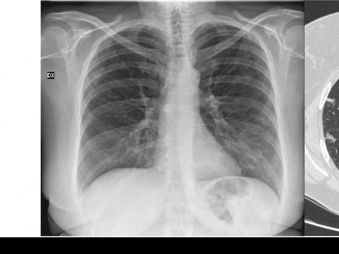
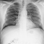
46-year-old asymptomatic female patient underwent chest radiography followed by CT since her husband was diagnosed of Covid-19 pneumonia (case n 14). pO2: 98%.
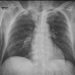
Chest radiography

Interstitial markings in the lower lobes with multiple ill-defined blurred consolidations. No pleural effusion.
CT


Multiple and bilateral consolidations at different stages, with predominant peribronchial and subpleural distribution. No pleural effusion. The patient was quarantined after performing the nasal swab.


